University of California, Davis

Category: ecology

The eyes of reef fishes

[cross-posted on my personal blog, as well]

Peter and I recently published a paper in BMC Evolutionary Biology and today the final HTML and PDF versions have become available. BMC is an open access journal, so everyone can read the paper:

Schmitz, L. & P.C. Wainwright (2011). Nocturnality constrains morphological and functional diversity in the eyes of reef fishes. BMC Evolutionary Biology, 11: 338. html, reprint.
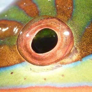
We have several really interesting results. The eye morphology of nocturnal reef teleosts is characterized by a syndrome that indicates good light sensitivity. Nocturnal fishes have large relative eye size, high optical ratio and large, rounded pupils. However, there is a trade-off. Improved optical light sensitivity comes at the cost of reduced depth of focus and reduction of potential accommodative lens movement. Diurnal reef fishes, which are released from the stringent functional requirements of vision in dim light, have much higher morphological and optical diversity than nocturnal species. Diurnal fishes have large ranges of optical ratio, depth of focus, and lens accommodation.

This paper is the first outcome of the analysis of a data set on the eye morphology of 265 species in 43 families of teleost reef fishes. It’s an enormous amount of data. All in all, we measured 5 traits in both eyes of 849 specimens, resulting in one of the largest data sets on eye morphology ever assembled. One aspect I would like to stress is that we measured eye morphology on fresh fish (in accordance to the UC Davis animal care protocol). The fixation process that preserved specimens went through may have altered eye morphology, so we wanted to avoid this potential problem.

I also would like to highlight one of the analyses in our paper. We assessed morphological diversity of diurnal and nocturnal species by calculating the combined variance of all shape axes of a principal component analysis. However, there are far more diurnal (n=211) than nocturnal species (n=54) in the data set, and ultimately the results may be biased because of uneven sampling. Inspired by a suggestion from David Bellwood we designed a simple rarefaction analysis. We randomly re-sampled 54 diurnal species (matching the number of nocturnal species) without replacement and calculated variance on PCs 2-5, and repeated this procedure 100, 000 times. This resulted in 100,000 PC analyses with the same number of diurnal and nocturnal species, with diurnal species randomly selected anew for each run. Then, we compared the distribution of nocturnal variances to the bootstrap distribution of diurnal variances. The results are convincing and, importantly, should no longer be biased by uneven sampling. The rarefaction is easily done in ‘R’; let me know if you are interested in the code.

Finally, here are thumbnails of all figures in the paper, with links to the corresponding to the high-resolution files. Feel free to use for teaching purposes.

Stickleback camouflage

This week, the Wainwright blog returns to a topic of perennial interest, the threespine stickleback. I will discuss a recent paper from the Schluter lab at UBC on color plasticity and background matching in stickleback.

To set the stage, it’s important to realize that from a stickleback’s perspective, “bird” is a four-letter word. Predation by diving birds like grebes and coots is commonplace in many freshwater stickleback populations. Unlike predatory dragonfly larva, which detect prey by vision and by water movement, diving birds generally detect their prey by sight alone. In other words, if you’re a freshwater stickleback, it’s very important that the top of your body blends in with your surroundings.

This stickleback didn't get the memo. (http://www.lifeontheslea.co.uk )

In this paper, Jason Clarke and Dolph Schluter tried to assay background matching capability between limnetic and benthic sticklebacks in Paxton Lake, British Columbia. First, they used a spectrometer to record the background color in the limnetic and benthic habitats. The open-water limnetic habitat was a bluish color, but the benthic habitat, which has more aquatic vegetation, tended to be more greenish. Additionally, the benthic habitat showed much more variation in color than the limnetic habitat.

After checking the background color, the authors painted two sets of cups, one designed to look like the limnetic background, and one designed to look like the benthic background. Then they put benthic and limnetic sticklebacks on each background, let them adjust their color for 15 minutes, photographed each fish, then measured how well each fish matched its background. They also did the same experiment again, but this time taking pictures every 20 seconds.

What did they find? Limnetic fish and benthic fish were equally good at matching the blue limnetic background, but limnetic fish were not as good at matching the green benthic background as benthics were. The time trial experiment helped to clear up what was going on: benthics rapidly adapted their colors to match the background, but limnetics were doing something different. Limnetic fish were cycling through different colors instead of fixing a particular color. Limnetics were more variable in color when viewed with a benthic background, but even on their “home turf” in the limnetic background, they still showed variation in color, but to a lesser degree.

The authors suggest that the patterns of color chance exhibited by benthics and limnetics are probably adaptive. Their spectrometer data indicates that the benthic habitat is more variable in color, and their background experiments show that benthics are better at rapidly changing their colors to match the background. The limnetic habitat, on the other hand, is much more uniform, so there would be little incentive for limnetics to evolve rapid color matching. However, limnetics may be adapting to their light environment in an entirely different way:  the  “flickering” exhibited by limnetics could be an adaptation to fluctuating light intensity in open water.

After reading this paper, I’m particularly curious what the color-matching abilities of the ancestral marine sticklebacks are like. If they resemble the limnetic, then this color matching ability will be another interesting benthic stickleback adaptation. It will be cool to see if it is possible to discern the genetic basis for this shift in plasticity.

Clark JM, Schluter D. Colour plasticity and background matching in a threespine stickleback species pair. Biological Journal of the Linnean Society. DOI: 10.1111/j.1095-8312.2011.01623.x

An optical illusion?

Zooplanktivory is one of the most distinct feeding niches in coral reef fish and many morphological traits have been interpreted as adaptations to feeding on plankton in the water column above the reef. One of these traditional hypotheses is that zooplanktivorous fish have larger eyes for sharper visual acuity. A larger eye usually has a longer focal length and thus is expected to produce a better-resolved image.

Peter and I tested this hypothesis with a data set on eye morphology of labrids (wrasses and parrot fishes):

Schmitz, L. & P.C. Wainwright (2011). Ecomorphology of the eyes and skull in zooplanktivorous labrid fishes. Coral Reefs, 30: 415-428. reprint.

Labrids are a species-rich clade of reef fish with enormous morphological and ecological diversity. We sampled a total of 21 species, with three independent origins of zooplanktivory: Clepticus parrae, the Creole Wrasse (photo: fishbase.org), Halichoeres pictus, the Rainbow Wrasse (photo: wetwebmedia.com), and Cirrhilabrus solorensis, the Red-eyed Fairy Wrasse (photo: fishbase.org).

To our surprise we failed to find any indication of larger eyes in zooplanktivores. We tried several methods, including phylogenetic residuals of eye diameter on body mass and evolutionary changes in eye size along branches leading to zooplanktivores, but zooplanktivorous labrids did not show any signs of having larger eyes than other trophic specialists. Instead, we suspect that the notion of large eyes in zooplanktivorous labrids is an optical illusion evoked by a size reduction of the anterior facial region, which makes the eye look bigger.

However, we did find other features interpreted as adaptations to zooplanktivory in labrids. Both Clepticus parrae and Halichoeres pictus have a large lens for given axial length of the eye, related to better visual acuity, a round pupil, possibly an adaptation to search a three-dimensional body of water for food, and longer gill rakers to help retain captured prey.

Our results are quite interesting in that they highlight the importance of many-to-one-mapping in form-function relations. There often is more than one possible pathway to perform a function. In labrids, increase in eye size to improve visual acuity apparently is not part of the evolutionary response. But, let’s see what we can find in other groups!

Nocturnal dinosaurs

Yet another non-teleost blog, but an interesting example of functional morphology and phylogenetic comparative methods. I’m cross-posting this on my own blog, too.

Nocturnal dinosaurs. Wait a second! Is that right? Nocturnal (= night-active) dinosaurs? Yes, indeed. Contrary to what was commonly believed, many dinosaurs were nocturnal.  We have to change our perception of the dinosaur era.

All details about methods, results, and implications can be found in these two siblings papers in Science and Evolution (thanks to AAAS and Wiley for being so extremely helpful in coordinating this release!):

L. Schmitz, R. Motani, Nocturnality in dinosaurs inferred from scleral ring and orbit morphology. Science, published online 14 April 2011 (10.1126/science.1200043)

R. Motani, L. Schmitz, Phylogenetic versus functional signals in the evolution of form-function relationships in terrestrial vision. Evolution, published online 14 April 2011 (10.1111/j.1558-5646.2011.01271)

We make several important points in these papers: (i) Dinosaurs were not restricted to day-activity (diurnality) by any means, (ii) Activity patterns evolve following ecological characteristics (e.g., diet+body size, terrestrial or flying), (iii) Physics of the environment drive the evolution of shape, (iv) We have a great new tool for reconstructing ecology of extinct species.

So, why did everyone think that dinosaurs were diurnal in the first place? We think there are at least two (historical) reasons. First, several decades ago dinosaurs were portrayed as sluggish, “cold-blooded” animals. It seemed too unlikely that these animals were capable of being active at night, when ambient temperatures were cold compared to the day. Second, the idea of diurnal dinosaurs may have arisen because many (paleo)biologists were trying to explain why the majority of mammals is nocturnal – and the picture of dinosaurs occupying the diurnal niche, pushing the mammals into the dark just seemed to fit all too well.

Well, that story isn’t all that simple and straight-forward anymore. Dinosaurs were nocturnal, too. And we don’t even know if the earliest mammals were actually nocturnal (Kenneth Angielczyk at the Field Museum and I are working on this right now). So, the assumed split in activity pattern between mammals and dinosaurs is certainly rejected. However, we do see a different pattern emerging. It appears as if the evolution of activity patterns is driven by ecology.

We found striking similarities between the Mesozoic and today’s biosphere. Large herbivores, just like living ‘megaherbivores’, were active both day and night, probably because of foraging needs (they just had to eat most of the time…), except for the hottest hours of the day when there was risk  of overheating.  Small carnivores such as Velociraptor were nocturnal hunters. Flying species, including early birds and pterosaurs (like Scaphognathus above, with scleral ring highlighted in blue color) were mostly day-active (although some of the pterosaurs were actually nocturnal). These ecological patterns are also found among today’s living mammals, lizards, and birds.

So, how can we tell? Eye shape is the key. Nocturnal animals roam around in low light during the night, and their eyes show characteristics that relate to improved light sensitivity. Eye soft-tissues rarely if ever fossilize, but many vertebrates (e.g., teleost fish, lizards, and birds) have an extra bone element within their eyes, the so called scleral ring. Notably, neither mammals, crocodiles, nor snakes have scleral rings [for more details I would refer you to the work of Tamara Franz-Odendaal]. The scleral ring, however, is present in basal archosaurs, pterosaurs, and dinosaurs, which enables us to reconstruct eye shape in these fossils..

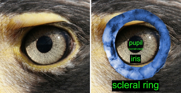
If you look at a bird eye (photos below) you will see the pupil opening defined by the iris. The scleral ring, situated within the sclera, the ‘leathery’ layer of the eye, surrounds the iris. The shape of the scleral ring and eye socket closely reflects eye shape, enabling us to distinguish nocturnal and diurnal dinosaurs.

For example, if you compare the scleral rings below, the nocturnal species on the right, a potoo, has a much larger ‘aperture’, or inner diameter than the diurnal hawk compared to eye socket size. Of course, the differences are often more subtle, so we use a Discriminant Analysis (DA) to retrieve a quantitative prediction. Prediction from a DA are rarely perfect, but overall the DA performs very well.

The relation between form and function of the eye is another convincing example how physics of the environment, that is light levels, drive the evolution of shape. However, it is not just physics that influences the evolution of shape. Shared ancestry is an important factor that may blur the form-function relation. Simply put, closely related species are expected to be more similar to each other than more distantly related species, so shape alone could potentially be misleading. With Ryosuke leading this part of the study, we solved this issue and developed a new method (implemented in ‘R’) that makes it possible to account for this phylogenetic bias. A step-by-step guide to this analysis is provided in the Supplementary Information of the paper in Science (www.sciencemag.org/cgi/content/full/science.1200043/DC1). We hope that this method will be helpful for other studies dealing with inferences of ecology from shape.

Eye shape revisited

In the anticipation of the publication of the dinosaur paper I thought it may be timely to highlight one of my latest papers. [Please note that this blog is also cross-posted on ecomorph.wordpress.com, my personal webpage].

Schmitz, L. & R. Motani (2010). Morphological differences between the eyeballs of nocturnal and diurnal amniotes revisited from optical perspectives of visual environments. Vision Research, 50: 936-946. [doi:10.1016/j.visres.2010.03.009]

This paper was one of the first steps towards inferring activity pattern in extinct dinosaurs and pterosaurs. We needed to find out whether it is possible to distinguish night-active and day-active living species before we could make any quantitative inferences in fossils. It was known previously that activity patterns indeed influence eye shape (see, for example, papers by Margaret Hall, Chris Kirk, and Callum Ross), but a reliable method to make quantitative predictions was still lacking.

Here is the short version of our approach: The eyes of land vertebrates cope with highly different light levels, largely depending on the preferred activity time of the animal. Night-active (nocturnal) species experience much lower light levels than day-active (diurnal) species. Twilight-active (crepuscular) and day-and-night-active species (cathemeral) species are exposed to both high and low light levels. The problem with low light levels is that it is very difficult to form a good image.

Overall, then, these contrasting lifestyles (and light levels) should drive the evolution of eye shape, because nocturnals need to have eyes with very good light sensitivity, whereas cathemerals and especially diurnals can optimize other aspects of vision.

We tackled this problem by applying Discriminant Analysis, focusing on features of macro-morphology that are correlated with optical function. Indeed, different activity patterns occupy distinct areas in morphospace and are identified with high accuracies (see the image to the left, modified from the Vision Res. paper). Diurnals are represented by open symbols, gray symbols are cathemerals, and black symbols are nocturnals.

The great news for paleobiology was that this method not only works with eye soft-tissue dimensions like eye diameter, axial length, or lens diameter (which rarely if ever fossilize), but also with skeletal features (i.e., size of the orbit and scleral ring) of birds. These skeletal structures are still correlated with optical function, even though their shape is influenced by other factors as well (see, for example, Schmitz 2009, J. Morph.). So, potentially one may be able to retrieve some or most of the optical information recorded in scleral ring and orbit structure… more on this next week!

Stickleblog: Spines hurt, according to predators

ResearchBlogging.orgOne of the distinguishing features of sticklebacks is that instead of having pelvic and dorsal fins, they have serrated bony spines that the fish can lock into place(more on the locking in a later entry).

Why would evolution result in a lineage of fishes that has spines instead of fins? The classic explanation is that spines make sticklebacks a painful meal; predators will avoid eating sticklebacks if other food is available.

In 1956, Hoogland et al tested whether stickleback spines were an effective defense against larger fish. The paper itself is 33 pages, with multiple experiments – for today’s entry, I’m going to concentrate on only two of these.

In the first experiment, pike were presented with three different types of fish: 12 threespine sticklebacks, 12 ninespine sticklebacks, and 12 carplike fish lacking spines. At first, the pike went after sticklebacks, with decidedly ouch-inducing results:

After eating one stickleback of each type, the pike focused exclusively on the fish without spines, eating all 12 of them in 5 days. Once all of these were gone, sticklebacks started disappearing, but at a much slower pace, with ninespine stickleback eaten faster than threespines. It’s difficult to conclude anything too comprehensively from this, as the authors didn’t do much in the way of replication, but it does suggest that fish predators prefer nonspined prey.

Then, the authors tried the obvious experiment – if threespine stickleback have spines that make it difficult for predators to eat them, what happens if the spines are removed? Once the spines were removed from a stickleback, predators stopped spitting them out and treated them similarly to the carplike fish.

Provided one is willing to overlook the paper’s archaic methodology and lack of rigorous statistical methods(and it is from the 1950s, remember), spines appear to decrease the deliciousness of stickleback.

Perhaps that’s why sticklebacks have never really taken off as a cuisine…

R. Hoogland, D. Morris and N. Tinbergen (1956). The Spines of Sticklebacks (Gasterosteus and Pygosteus) as Means of Defence against Predators (Perca and Esox) Behaviour, 10 (3), 205-236 DOI: 10.2307/4532857

The ENMTools web site is getting ready for launch

For anyone out there who read our Evolution paper last year, Rich Glor, Michael Turelli, and I are putting together a web site to host the software we made for that study. It’s got a bunch of other little bits and bobs in development as well, mostly revolving around different resampling procedures for use with environmental niche modeling. You can find it at www.enmtools.com. I’ll post any major developments here as well.

Yes, the site is supposed to look that way.

Datamuncher – a handy tool for niche and distribution modelers

Here’s a little tool I whipped together for my own use. I hope it’s useful to others as well. Basically what it does is take .csv files of species occurrences and a batch of ASCII files, and converts them into three output files. They are:

  1. A community presence/absence matrix, with “community” defined as a grid cell in the ASCII raster files.
  2. A set of coordinates for each grid cell that has at least one species present in it, corresponding to the “communities” above.
  3. A matrix of values from the ASCII raster files for each community.

The general idea is to take data in a format that is acceptable for Maxent (.csv and .asc) and convert it into a set of files that are usable with some of the R algorithms. Currently it seems to be working with GDM, but I haven’t tried anything else. You may need to delete columns 1-3 in the environment file, depending on what you’re doing with it.

Here’s the quick and dirty of it:

Where it says “occurrence files”, you just hit the “add files” button and drop in all of the .csv files you want to use. Note that everything in that box is going to be thrown into ONE set of output files!

Second, you add your environmental layers. ASCII raster format is the only one it understands.

Third, pick an output directory.

Finally, name your analysis. If your analysis is named “my_stuff”, the output files will be “my_stuff_communities.csv” (1 above), “my_stuff_community_coordinates.csv” (2), “my_stuff_environment.csv” (3), all dropped into the output directory you chose.

In addition to its original intent, this tool may also be useful to those who want a quick and dirty way to get their data extracted for a MANOVA or other analysis – if you feed it occurrence files for one species at a time, the environment file will contain all conditions occupied by that species. Neat!

You can download it as a Windows executable file here, or as a Perl script>here. Be aware that the Perl script requires that Tk be installed (NOT Tk+!), which you can do from the package manager. Also be aware that it probably won’t work on a Mac because of the way it’s parsing directories. If anyone wants a Mac version, please feel free to email me.

Also let me know if you hit any snags. Testing has been rather limited so far, as I just finished it today. Use at your own risk!

© 2025 Wainwright Lab

Theme by Anders NorenUp ↑