University of California, Davis

Category: Uncategorized (Page 1 of 2)

Steps towards understanding comparative methods

Using phylogenetic comparative methods warrants a basic understanding of the history and progress of this field.  Working with some of the more recent tools for comparative evolutionary biology, I feel compelled to find out how current methods were devised, whom to credit for the methods I use, and what assumptions I am making by using them.  Below is a list of some of the landmark papers in comparative methods, with comments and synopses (written by me and Tomomi).

Felsenstein (1981) describes the basics for creating a maximum likelihood tree from a set of nucleotide sequences. One step elaborated from his 1973 paper is Felsenstein’s pruning algorithm for calculating the likelihood of a phylogenetic tree given branch lengths and tip values.  This algorithm makes likelihood calculations more computationally efficient by eliminating redundant calculations.  The paper also describes the Markov process for finding the maximum likelihood tree from nucleotide data.  Felsenstein uses a substitution model for molecular phylogenetics in which each nucleotide has a different stationary frequency (A, C, G, and T are not expected to be equally represented at any given site on DNA sequences).  Methods for searching tree space have been improved, and Bayesian theory has since permeated phylogenetic analyses, but the pruning algorithm continues to be an important subroutine in phylogenetic computations.

Possibly the most cited paper in phylogenetic comparative methods, Felsenstein (1985) describes with clear examples why species trait values may not be statistically independent and what might be done to compensate.  Felsenstein elaborates on his method of calculating standardized contrasts (phylogenetically independent contrasts) to help overcome the non-independence of character traits.  These contrasts are basically the differences between trait values of species pairs weighted by the evolutionary change separating them; they are estimates of the rate of change over time. A common use of standardized contrasts is to look for correlation in this rate between two traits; if standardized contrasts of traits X and Y are compared in a regression analysis, a linear trend suggests correlated rates of evolution between the traits.

Schluter et al. (1997) discuss the need for error estimates on ancestral state reconstructions. The paper introduces maximum likelihood ancestral state reconstructions of both discrete and continuous characters.   Responding to Schluter et al.’s call to account for error in tree construction, Huelsenbeck et al. (2003) describe a Bayesian method for mapping the change in character states onto a phylogeny.  The introduction reiterates the importance of having an alternative to parsimony methods when tackling character change; as with maximum likelihood, the new methods allow for more than one change along a given branch in the tree.  While the Huelsenbeck et al. paper is a landmark for evolutionary analysis, it also contains a very coherent introduction to the instantaneous rate matrix, substitution model, and likelihood calculations for finding the probabilities of evolutionary histories.

Although Brownian motion is often used to model quantitative character evolution, the Ornstein-Uhlenbeck (OU) process can also be used to develop informative evolutionary models.  OU models incorporate selection as a selective optima, or adaptive peak.  OU and Brownian motion are not entirely unrelated as OU collapses to Brownian motion in the absence of selection.  Butler and King (2004) use OU to test which of several evolutionary models has the best fit to several example data sets involving anoles.  They use likelihood ratio tests to determine how various OU-based models perform against Brownian motion, observing that biological information is important in determining what models to consider.  They also stress that stasis, although positive support for stabilizing selection, is often disregarded and can lead to underestimation of evolutionary drift.  However, although they state that Brownian motion is a pure drift process, Brownian motion can provide a good fit to selective schemes under fluctuating directional selection (O’Meara et al. 2006).

While Grafen (1989) first describes a generalized least squares model for phylogenetic regressions, Chris O. from our lab pointed out the appendix of a paper on mammal intestines (Lavin et al. 2008) as a nice summary of PGLS.  This appendix describes both the history and the methods of phylogenetic regression analysis.  PGLS can be performed not only under a Brownian motion model, but also with OU and several other transformations of the variance-covariance matrix used in the mixed model approach.

These are just a handful of important papers; feel free to add to the list via comments with other references and their contributions.

Where do they get all those wonderful videos?

This blog is also posted on my personal blog.

I joined the Wainwright lab in October of last year. While I had experience with swimming fish, including high-speed video analyses, I had not done any filming of fish feeding. At the beginning of this year I got my first taste of obtaining high-speed videos of fish suction feeding. Since that time I have been amazed at the diversity of fish the lab studies (for example, check out the Inimicus didactylus video), the speed of the strikes, and kinematics during the strike; some of the little fish have quite a big gape to capture their prey. The data we are gathering is allowing us to get a glimpse of the patterns of diversity in the kinematics during suction feeding among various species of marine fish, as well as the potential morphological and mechanical correlates of that kinematic data.

Many of the videos that we obtain as a result of this research we upload to our Youtube channel to share with the public, usually the best videos, in focus and lateral. When we film we always try to get focused and lateral sequences for subsequent digitizations. These clear lateral videos allow us to digitize several landmarks on the fish during the strike sequence to get several kinematic variables such as maximum gape, time to pre capture, and ram speed to name a few. But we don’t just need clear lateral videos to showcase on Youtube; we mainly need clear videos to be able to track the landmarks throughout the sequence, and we need lateral videos to obtain accurate kinematics. For example, if the sequences are not clear, it may be harder to track a landmark and there may be more error because the points may drift. If the fish isn’t completely lateral, we may not be able to see all the points, or if the fish as at an angle (going into the third dimension, such as toward the back of the aquarium, which we don’t capture in the 2-dimensional video) the kinematic variables may not be accurate. So, there is a reason for us obtaining these clear lateral videos. However, we also recognize that some of these strike sequences are pretty amazing, so we share them on Youtube.

Lately, our videos (especially the Inermia vittata video you can see in a previous post) have attracted the attention of several science, news and tech blogs. Thank you to all that have posted our videos. However, obtaining these videos is not always easy work, something else that I have learned since being a part of the Wainwright lab. Obtaining these sequences can sometimes (and often) take lots of hours of filming, patience, and hard work. Much of this depends on the fish or the species. Some fish are very good performers, and obtaining several good sequences does not take long (for example the Histrio histrio you can in another previous post). Others require some training to get the fish use to the lights required to capture the sequences at 1000 frames per second. Furthermore, not every fish feeds perfectly lateral every time, or we have multiple individuals in the aquarium that all want food, and the fish themselves are not always perfect. In fact, there are plenty of instances when the predator will miss the prey. This itself is interesting; a former Wainwright Lab member Tim Higham has done some work on the accuracy of strikes, what makes a predator accurate and what can make them miss? Perhaps having a farther strike distance and faster strike velocity decreases accuracy, but to compensate, species have larger gapes to ingest a greater amount of water to increase chances of prey capture (e.g., Higham et al. 2007). We recently posted a video on our Youtube channel of some of these ‘outtakes.’ Again, it is not always easy to capture the clear lateral videos and it takes a lot of work, so this video highlights a ‘bad day at the office’.

Patrick Fuller on feeding duty, Tomomi Takada on camera duty during a typical filming session.

So how do we get all these wonderful videos? First, it is almost always a two person job (although Matt has filmed sticklebacks alone). One person feeds the fish, trying to get them in view of the camera, and striking laterally. This job is almost an art form in itself. You have to learn the behavior of the fish; are they sit and wait predators like the frogfish, fast strikers like the white-streaked grouper, or more active swimmers like Inermia vittata? Therefore, the person feeding has to be aware of the fish’s behavior to try and get good sequences. Challenges may also arise is there are multiple individuals.

Another view of a filming session

We want to ensure all fish eat and we want to get sequences from all individuals, so the person feeding has to keep track of the fish or target the various individuals. The other person involved in the process is the person responsible for tracking the prey and predator, focusing the camera and triggering the high-speed camera. This job is also not easy. It takes some skill to track and focus and quickly trigger the camera. We film at 1000 frames per second and many of the videos on Youtube are played back at 10 frames per second. So what do these strikes actually look like in real time, how much time does the person manning the camera have to respond? To demonstrate this we made a video of a full sequence captured during filming,  in real time and about 200ms of that sequence played back at 10 frames per second for comparison. The person on camera duty has 3 seconds to trigger. You can see from the video, the person responsible for this part of filming either has, or hopefully obtains quick reflexes!

Although our Youtube channel features some of the best sequences we capture, keep in my mind we always strive to get the best videos. And the next time you see one of our videos on Youtube or elsewhere remember that one video is probably the product of hours of work. I want to also note that many of these videos are the work of undergraduate assistants we have in the lab. Many of our Youtube ‘stars’ were captured by our undergraduates, their assistance has been greatly appreciated and many of these videos would not have been captured without them.

Stickleback attack (part 1)

Since our last video posting, many of the videos on our lab’s Youtube channel have gone viral. As of this blog post, the video of Inermia vitatta has accrued over 120,000 hits and has been featured on TV programs and newspaper articles around the globe. Not bad for a small fish!

[youtube=http://www.youtube.com/watch?v=psdLbN7skg4]

Today’s video features the threespine stickleback, Gasterosteus aculeatus, feeding on a cladoceran (Daphnia pulex). If you have a short attention span like me, one of the first things you’ll notice from the video is how shiny the fish is. The reflective armor plates and large spines are a clue that this is a threespine stickleback from an anadromous population. Anadromous stickleback have a life history similar to a miniature salmon – they are born in freshwater, travel to the ocean, then return to freshwater to breed. Unlike salmon, anadromous stickleback do not necessarily return to their home stream to breed. Anadromous stickleback also look very similar to each other – an Alaska anadromous fish looks very similar to a California anadromous fish.

Sometimes, these anadromous stickleback will travel to a newly-formed lake or river, and instead of returning to the ocean, some fish will stay in freshwater, founding a new population of freshwater stickleback. Over time, this freshwater population will evolve to better match its new freshwater habitat.

These anadromous and freshwater populations are one of the reasons stickleback are such a good system for studying evolutionary biology. We can study the result of rapid evolution in the freshwater populations, and then turn around and study the anadromous fish that resemble the fish that founded the freshwater population. Studying ancestral and derived populations is one of the few ways – short of a time machine – that we can learn the dynamics of adaptation in natural populations.

If we study how this anadromous stickleback captures prey, and then study how freshwater stickleback catch prey, we can learn a lot about the process of adaptation. I’ve devoted much of my PhD work to studying this system, and I’ll be talking more about it in future posts.

Stickleback camouflage

This week, the Wainwright blog returns to a topic of perennial interest, the threespine stickleback. I will discuss a recent paper from the Schluter lab at UBC on color plasticity and background matching in stickleback.

To set the stage, it’s important to realize that from a stickleback’s perspective, “bird” is a four-letter word. Predation by diving birds like grebes and coots is commonplace in many freshwater stickleback populations. Unlike predatory dragonfly larva, which detect prey by vision and by water movement, diving birds generally detect their prey by sight alone. In other words, if you’re a freshwater stickleback, it’s very important that the top of your body blends in with your surroundings.

This stickleback didn't get the memo. (http://www.lifeontheslea.co.uk )

In this paper, Jason Clarke and Dolph Schluter tried to assay background matching capability between limnetic and benthic sticklebacks in Paxton Lake, British Columbia. First, they used a spectrometer to record the background color in the limnetic and benthic habitats. The open-water limnetic habitat was a bluish color, but the benthic habitat, which has more aquatic vegetation, tended to be more greenish. Additionally, the benthic habitat showed much more variation in color than the limnetic habitat.

After checking the background color, the authors painted two sets of cups, one designed to look like the limnetic background, and one designed to look like the benthic background. Then they put benthic and limnetic sticklebacks on each background, let them adjust their color for 15 minutes, photographed each fish, then measured how well each fish matched its background. They also did the same experiment again, but this time taking pictures every 20 seconds.

What did they find? Limnetic fish and benthic fish were equally good at matching the blue limnetic background, but limnetic fish were not as good at matching the green benthic background as benthics were. The time trial experiment helped to clear up what was going on: benthics rapidly adapted their colors to match the background, but limnetics were doing something different. Limnetic fish were cycling through different colors instead of fixing a particular color. Limnetics were more variable in color when viewed with a benthic background, but even on their “home turf” in the limnetic background, they still showed variation in color, but to a lesser degree.

The authors suggest that the patterns of color chance exhibited by benthics and limnetics are probably adaptive. Their spectrometer data indicates that the benthic habitat is more variable in color, and their background experiments show that benthics are better at rapidly changing their colors to match the background. The limnetic habitat, on the other hand, is much more uniform, so there would be little incentive for limnetics to evolve rapid color matching. However, limnetics may be adapting to their light environment in an entirely different way:  the  “flickering” exhibited by limnetics could be an adaptation to fluctuating light intensity in open water.

After reading this paper, I’m particularly curious what the color-matching abilities of the ancestral marine sticklebacks are like. If they resemble the limnetic, then this color matching ability will be another interesting benthic stickleback adaptation. It will be cool to see if it is possible to discern the genetic basis for this shift in plasticity.

Clark JM, Schluter D. Colour plasticity and background matching in a threespine stickleback species pair. Biological Journal of the Linnean Society. DOI: 10.1111/j.1095-8312.2011.01623.x

An optical illusion?

Zooplanktivory is one of the most distinct feeding niches in coral reef fish and many morphological traits have been interpreted as adaptations to feeding on plankton in the water column above the reef. One of these traditional hypotheses is that zooplanktivorous fish have larger eyes for sharper visual acuity. A larger eye usually has a longer focal length and thus is expected to produce a better-resolved image.

Peter and I tested this hypothesis with a data set on eye morphology of labrids (wrasses and parrot fishes):

Schmitz, L. & P.C. Wainwright (2011). Ecomorphology of the eyes and skull in zooplanktivorous labrid fishes. Coral Reefs, 30: 415-428. reprint.

Labrids are a species-rich clade of reef fish with enormous morphological and ecological diversity. We sampled a total of 21 species, with three independent origins of zooplanktivory: Clepticus parrae, the Creole Wrasse (photo: fishbase.org), Halichoeres pictus, the Rainbow Wrasse (photo: wetwebmedia.com), and Cirrhilabrus solorensis, the Red-eyed Fairy Wrasse (photo: fishbase.org).

To our surprise we failed to find any indication of larger eyes in zooplanktivores. We tried several methods, including phylogenetic residuals of eye diameter on body mass and evolutionary changes in eye size along branches leading to zooplanktivores, but zooplanktivorous labrids did not show any signs of having larger eyes than other trophic specialists. Instead, we suspect that the notion of large eyes in zooplanktivorous labrids is an optical illusion evoked by a size reduction of the anterior facial region, which makes the eye look bigger.

However, we did find other features interpreted as adaptations to zooplanktivory in labrids. Both Clepticus parrae and Halichoeres pictus have a large lens for given axial length of the eye, related to better visual acuity, a round pupil, possibly an adaptation to search a three-dimensional body of water for food, and longer gill rakers to help retain captured prey.

Our results are quite interesting in that they highlight the importance of many-to-one-mapping in form-function relations. There often is more than one possible pathway to perform a function. In labrids, increase in eye size to improve visual acuity apparently is not part of the evolutionary response. But, let’s see what we can find in other groups!

Waiting in the weeds: the Sargassum frogfish (Histrio histrio)

This blog is cross-posted on my personal website’s blog.

This week’s blog will focus on one of the things the Wainwright Lab does best, fish suction feeding. One of the current projects in the lab is comparing the kinematics of suction feeding from a large inter-specific group of fish. To do this, we film fish feeding on various prey (mostly zebrafish) with a high-speed video camera at a 1000 frames per second. Once the sequence has been captured the videos are digitized to obtain several kinematic parameters associated with suction feeding, such as gape and jaw protrusion. A product of this research is some amazing videos of a diversity of fish using suction to subdue prey. Throughout this blog I will post some examples of the fish we have filmed. Hopefully these videos will allow you to observe not only the diversity in fish species, but also the diversity in the kinematics associated with suction feeding.

The first video comes from a fish that made its debut at UC Davis’ 97th annual Picnic Day this last Saturday. The Wainwright lab had a small demonstration for the public, showing some videos of the various fish in the lab. Also present were two charismatic frogfish on display for the public. The public got to see the live fish and their feeding kinematics in slow motion from some videos. One of the fish on display was the sargassum frogfish (Histrio histrio) of the family Antennariidae. One of the unique features of Antennariidae is that the first dorsal fin ray is modified into a lure to attract prey. These fish tend to be sit-and-wait predators so the lure helps bring in food.

The sargassum frogfish can be found in most tropical seas living among the Sargassum weeds, hence its name; often near reefs. It is the only frogfish with a swim bladder, allowing it to float among the weeds. They can reach a size of 20 cm total length (from the most anterior tip to the edge of the caudal fin); and their color patterning is a mottled greenish/brown with numerous weedy dermal appendages. This patterning allows the sargassum frogfish to blend in to the weeds waiting for unsuspecting fish or shrimp. Unlike some of the other frogfish, this species swims more in open water, and some divers have noted their ability to jump out onto the floating weeds to avoid potential threats.

In terms of their feeding, these fish are mainly sit-and-wait predators, hiding among the weeds; however they do have the ability to swim after prey.  The lure in this species is small compared to some other frogfish.  Once the prey is close enough, the frogfish initiates its strike using suction to obtain the prey. Being a sit-and-wait predator these frogfish have little to no body displacement (you can see it is not swimming to capture the prey item in the video below), but you may also notice their mouth can displace a good distance in order to capture the prey. This clip is a mid-water strike, typical of this species. These strikes are also extremely quick. This video was filmed at 1000 frames per second, but is playing back at 10 frames per second; so the actual strike is much faster than the video playback.  One of the things we are interested in is the variation in kinematics of suction feeding associated with different ecologies. Have sit-and-wait predators evolved a greater mouth displacement compared to more active foragers? The data we are collecting will be able to shed light on some of these questions, and the sargassum frogfish is a good example of a sit-and-wait predation strategy.

Nocturnal dinosaurs

Yet another non-teleost blog, but an interesting example of functional morphology and phylogenetic comparative methods. I’m cross-posting this on my own blog, too.

Nocturnal dinosaurs. Wait a second! Is that right? Nocturnal (= night-active) dinosaurs? Yes, indeed. Contrary to what was commonly believed, many dinosaurs were nocturnal.  We have to change our perception of the dinosaur era.

All details about methods, results, and implications can be found in these two siblings papers in Science and Evolution (thanks to AAAS and Wiley for being so extremely helpful in coordinating this release!):

L. Schmitz, R. Motani, Nocturnality in dinosaurs inferred from scleral ring and orbit morphology. Science, published online 14 April 2011 (10.1126/science.1200043)

R. Motani, L. Schmitz, Phylogenetic versus functional signals in the evolution of form-function relationships in terrestrial vision. Evolution, published online 14 April 2011 (10.1111/j.1558-5646.2011.01271)

We make several important points in these papers: (i) Dinosaurs were not restricted to day-activity (diurnality) by any means, (ii) Activity patterns evolve following ecological characteristics (e.g., diet+body size, terrestrial or flying), (iii) Physics of the environment drive the evolution of shape, (iv) We have a great new tool for reconstructing ecology of extinct species.

So, why did everyone think that dinosaurs were diurnal in the first place? We think there are at least two (historical) reasons. First, several decades ago dinosaurs were portrayed as sluggish, “cold-blooded” animals. It seemed too unlikely that these animals were capable of being active at night, when ambient temperatures were cold compared to the day. Second, the idea of diurnal dinosaurs may have arisen because many (paleo)biologists were trying to explain why the majority of mammals is nocturnal – and the picture of dinosaurs occupying the diurnal niche, pushing the mammals into the dark just seemed to fit all too well.

Well, that story isn’t all that simple and straight-forward anymore. Dinosaurs were nocturnal, too. And we don’t even know if the earliest mammals were actually nocturnal (Kenneth Angielczyk at the Field Museum and I are working on this right now). So, the assumed split in activity pattern between mammals and dinosaurs is certainly rejected. However, we do see a different pattern emerging. It appears as if the evolution of activity patterns is driven by ecology.

We found striking similarities between the Mesozoic and today’s biosphere. Large herbivores, just like living ‘megaherbivores’, were active both day and night, probably because of foraging needs (they just had to eat most of the time…), except for the hottest hours of the day when there was risk  of overheating.  Small carnivores such as Velociraptor were nocturnal hunters. Flying species, including early birds and pterosaurs (like Scaphognathus above, with scleral ring highlighted in blue color) were mostly day-active (although some of the pterosaurs were actually nocturnal). These ecological patterns are also found among today’s living mammals, lizards, and birds.

So, how can we tell? Eye shape is the key. Nocturnal animals roam around in low light during the night, and their eyes show characteristics that relate to improved light sensitivity. Eye soft-tissues rarely if ever fossilize, but many vertebrates (e.g., teleost fish, lizards, and birds) have an extra bone element within their eyes, the so called scleral ring. Notably, neither mammals, crocodiles, nor snakes have scleral rings [for more details I would refer you to the work of Tamara Franz-Odendaal]. The scleral ring, however, is present in basal archosaurs, pterosaurs, and dinosaurs, which enables us to reconstruct eye shape in these fossils..

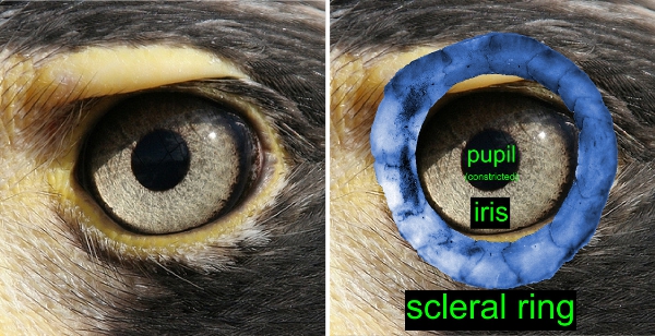
If you look at a bird eye (photos below) you will see the pupil opening defined by the iris. The scleral ring, situated within the sclera, the ‘leathery’ layer of the eye, surrounds the iris. The shape of the scleral ring and eye socket closely reflects eye shape, enabling us to distinguish nocturnal and diurnal dinosaurs.

For example, if you compare the scleral rings below, the nocturnal species on the right, a potoo, has a much larger ‘aperture’, or inner diameter than the diurnal hawk compared to eye socket size. Of course, the differences are often more subtle, so we use a Discriminant Analysis (DA) to retrieve a quantitative prediction. Prediction from a DA are rarely perfect, but overall the DA performs very well.

The relation between form and function of the eye is another convincing example how physics of the environment, that is light levels, drive the evolution of shape. However, it is not just physics that influences the evolution of shape. Shared ancestry is an important factor that may blur the form-function relation. Simply put, closely related species are expected to be more similar to each other than more distantly related species, so shape alone could potentially be misleading. With Ryosuke leading this part of the study, we solved this issue and developed a new method (implemented in ‘R’) that makes it possible to account for this phylogenetic bias. A step-by-step guide to this analysis is provided in the Supplementary Information of the paper in Science (www.sciencemag.org/cgi/content/full/science.1200043/DC1). We hope that this method will be helpful for other studies dealing with inferences of ecology from shape.

Eye shape revisited

In the anticipation of the publication of the dinosaur paper I thought it may be timely to highlight one of my latest papers. [Please note that this blog is also cross-posted on ecomorph.wordpress.com, my personal webpage].

Schmitz, L. & R. Motani (2010). Morphological differences between the eyeballs of nocturnal and diurnal amniotes revisited from optical perspectives of visual environments. Vision Research, 50: 936-946. [doi:10.1016/j.visres.2010.03.009]

This paper was one of the first steps towards inferring activity pattern in extinct dinosaurs and pterosaurs. We needed to find out whether it is possible to distinguish night-active and day-active living species before we could make any quantitative inferences in fossils. It was known previously that activity patterns indeed influence eye shape (see, for example, papers by Margaret Hall, Chris Kirk, and Callum Ross), but a reliable method to make quantitative predictions was still lacking.

Here is the short version of our approach: The eyes of land vertebrates cope with highly different light levels, largely depending on the preferred activity time of the animal. Night-active (nocturnal) species experience much lower light levels than day-active (diurnal) species. Twilight-active (crepuscular) and day-and-night-active species (cathemeral) species are exposed to both high and low light levels. The problem with low light levels is that it is very difficult to form a good image.

Overall, then, these contrasting lifestyles (and light levels) should drive the evolution of eye shape, because nocturnals need to have eyes with very good light sensitivity, whereas cathemerals and especially diurnals can optimize other aspects of vision.

We tackled this problem by applying Discriminant Analysis, focusing on features of macro-morphology that are correlated with optical function. Indeed, different activity patterns occupy distinct areas in morphospace and are identified with high accuracies (see the image to the left, modified from the Vision Res. paper). Diurnals are represented by open symbols, gray symbols are cathemerals, and black symbols are nocturnals.

The great news for paleobiology was that this method not only works with eye soft-tissue dimensions like eye diameter, axial length, or lens diameter (which rarely if ever fossilize), but also with skeletal features (i.e., size of the orbit and scleral ring) of birds. These skeletal structures are still correlated with optical function, even though their shape is influenced by other factors as well (see, for example, Schmitz 2009, J. Morph.). So, potentially one may be able to retrieve some or most of the optical information recorded in scleral ring and orbit structure… more on this next week!

Catch Peter on National Geographic TV tonight!

If you’re quick you might be able to catch Peter on National Geographic TV tonight on the show “Hooked On Fish”. It’s a great show, using stories of anglers catching huge fish as a jumping-off point to talk about some fascinating natural history and conservation issues.

If you’re not fast enough to see it on the air tonight, you can at least get a sample of it here.

Dechronization interviews Joe Felsenstein

Just in case anyone is reading this blog who is not also reading Dechronization, I have two things to say. First, what is wrong with you? Second, Luke Harmon and Dan Rabosky just posted a great interview of Joe Felsenstein, which you should read. If you don’t know who that is, see point 1 above. You can find the interview here.

« Older posts

© 2024 Wainwright Lab

Theme by Anders NorenUp ↑